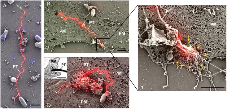
Fig 2. Correlative light and electron microscopy (CLEM) analysis of E. hellem infection.
E. hellem infected host cells were initially incubated with rab-PcAb-EhPTP1 (rabbit polyclonal antibody; red) and MAb-EhPTP4 (mouse monoclonal antibody; green). The fluorescence image and SEM image of same sites were taken sequentially, and the fluorescence images of EhPTP4 and the polar tube were correlated to the SEM images. Panel A shows a germinated polar tube with the EhPTP4 staining at the end of tube (white arrow). Panel B shows the binding of polar tube to host cell plasma membrane during infection. Panel C is an enlarged section of panel B, the tip of polar tube and some host fiber-like structures (yellow arrows). Panel D demonstrates penetration of polar tube into host cell plasma membrane, there was no EhPTP4 staining at the end of polar tube (white arrow), suggested that the end of polar tube had already entered the host cell and was below the level of imaging. Panel E shows TEM data demonstrating that the polar tube was surrounded with host cell membrane (black arrows) at the site of infection. PT, polar tube; PM, plasma membrane. Bar size: 2 μm panels A, B, C and D, and 400 nm panel E.

