Figure 1.
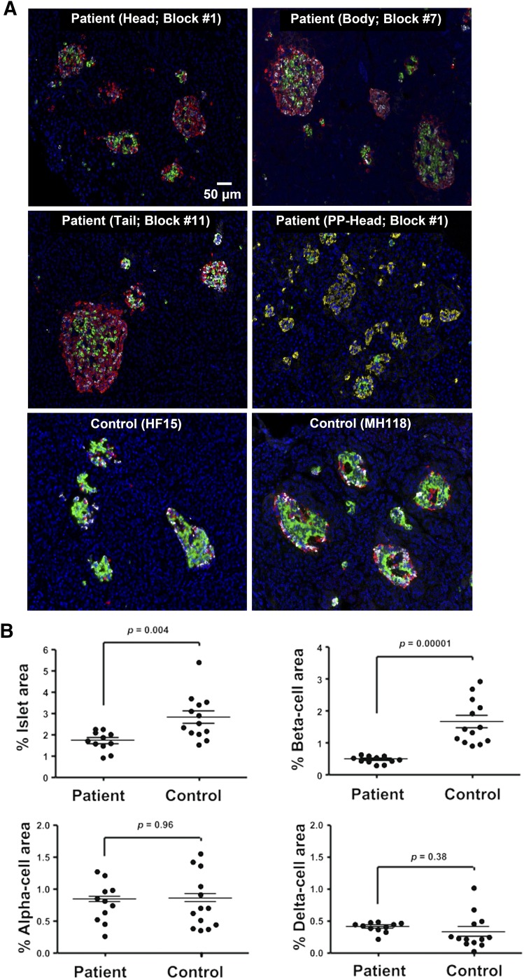
Histology in a 2-year-old female patient with KCNJ11 mutation p.G334D. (A) Representative islets from the head, body, and tail regions of the pancreas from the patient and two representative age-matched control subjects stained for insulin (green), glucagon (red), somatostation (white), and 4′,6-diamidino-2-phenylindole (blue). An additional panel (PP-Head) from the patient is stained for the pancreatic polypeptide (yellow), insulin (green), glucagon (white), and 4′,6-diamidino-2-phenylindole (blue). (B) Endocrine cell area. The percentage of pancreatic area that is islets, β-cells, α-cells, and δ-cells is shown for all 11 blocks in the patient and one section from each of the 13 age-matched control subjects. Horizontal lines represent mean ± SEM.

