Figure 2.
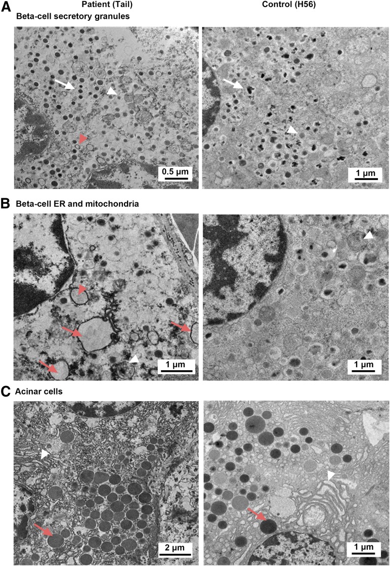
EM analysis. (A) EM ultramicrograph of a β-cell from patient and control showing the morphology of the secretory granules. Ultrastructural analysis revealed altered morphology of β-cells in the patient with immature granules (light dense core, white arrowhead), empty granules (red arrowhead), and reduced crystalline cores in the patient (white arrow) compared with the control. Note that nuclei showed normal morphology in both panels. (B) Endoplasmic reticulum and mitochondria structure in patient and control. A deteriorated β-cell contained several dilated rough endoplasmic reticulum (ER, red arrow) that were filled with flocculent material (presumably protein). One close to a nucleus (red arrowhead) appears to be directly attached to a perinuclear membrane and an underlying perinuclear space, likely to represent the conduit of protein production. A partially degenerated mitochondrion is seen in the patient (white arrowhead), whereas all mitochondria in the control appear normal (white arrowhead). (C) Pancreatic acinar tissue in patient and control. Zymogen granules (red arrow) appear normal in both patient and control.

