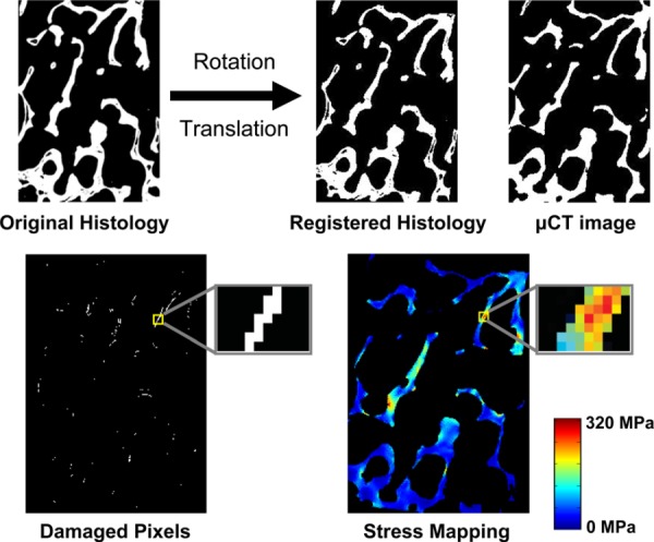
Fig. 4.

Example of two-dimensional automated image registration (top). Histology section is iteratively rotated and translated to optimally align with the micro-CT section. Once registered, the pixel-by-pixel method was used to analyze the stresses/ strains in microdamaged and undamaged pixels (bottom).
