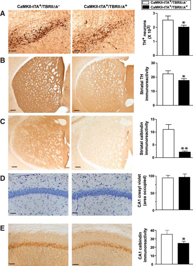
Figure 3.

Mice with reduced TGF-β signaling in neurons display degeneration of the nigrostriatal system. CaMKII-tTA+/TBRIIΔk+ mice were generated by crossing CaMkII-tTA mice with tet-O-TBRIIΔk mice expressing a kinase-deficient TGF-β type II receptor. CaMKII-tTA+/TBRIIΔk− mice were used as controls. A, B, Brain sections from these mice were immunostained with an antibody against TH and were subjected to stereological analysis of TH+ DA neurons in substantia nigra pars compacta (A) and TH+ nerve terminals in the striatum (B). C, E, Brain sections were immunostained with an antibody against calbindin, and calbindin immunoreactivity was measured in the striatum (C) and hippocampal CA1 (E). D, Brain sections were stained with 0.02% Cresyl Violet. Scale bar, 50 μm. Bars represent the mean ± SEM and were analyzed by unpaired t test. *p < 0.05; **p < 0.01. n = 10–12 mice/group.
