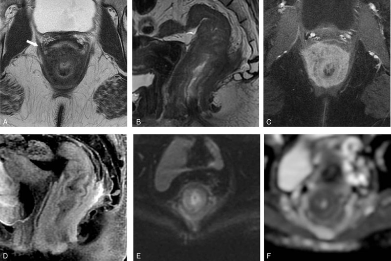
Figure 3.

On MRI rectum, noncontrast axial and sagittal T2-weighted images (TR/TE = 2440/82 ms; slice thickness = 4 mm; field of view = 18 cm) (A and B) show a long segment of concentric rectal wall thickening extending to the anal canal, with thickening of the various layers. The mural thickening involves the submucosa and muscularis propria but spares the mucosa. A T2 hypointense, extramural component (arrow) is also seen. Postcontrast axial and sagittal T1-weighted fat saturated image show avid enhancement of the rectal wall (C and D). Axial diffusion-weighted imaging (b = 800 s/mm2) and apparent diffusion coefficient (ADC) maps (E and F) show mild restricted diffusion of the thickened rectal wall (ADC = 1.1 × 10⁻³ mm2/s).
