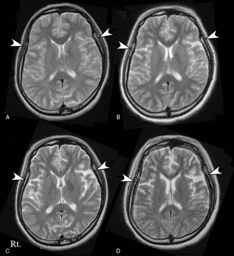
Figure 2.

Serial MRI studies in Patient 2. The first brain MRI was normal (panel A). The second brain MRI, obtained on day 19, showed the presence of diffuse brain atrophy (panel B, white arrows). The diffuse brain atrophy showed progression on the third brain MRI, obtained on day 42 (panel C), and the final follow-up MRI, obtained 3 years 8 months after admission (panel D).
