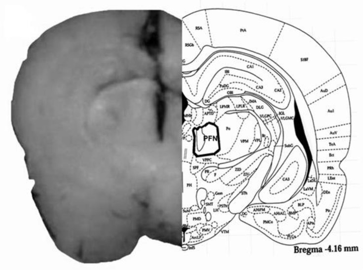
Fig. 1.
Schematic illustration of coronal section of the rat brain showing the approximate location of PFN microinjection sites in the experiments. Location of the injection cannulas tip in PFN (left side) of all rats was included in the data analysis. Atlas plate (right side) is adopted with permission from Paxinos and Watson

