Figure 7.
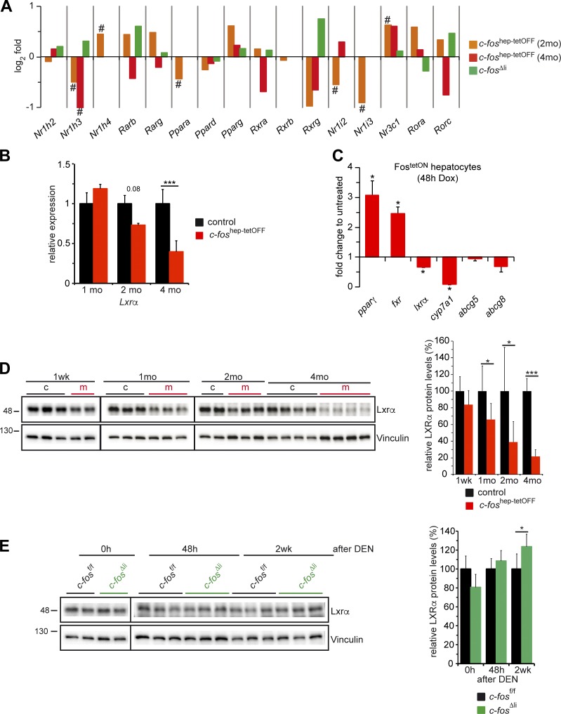
Regulation of LXRα expression by c-Fos. (A) Relative expression of the indicated nuclear receptors, including Nr1h3 encoding for LXRα, by RNA-seq in c-foshep-tetOFF mice at 2 and 4 mo of ectopic c-Fos expression; (n = 2; 3/cohort) and in c-fosΔli mice 48 h after DEN (n = 3/cohort). Bar graphs represent mean fold changes (log2); # indicates significance after multiple testing corrections. (B) qRT-PCR analyses of Lxrα in total liver tissue of c-foshep-tetOFF and control mice at 1, 2, and 4 mo of c-Fos expression. Mean expression in controls set to 1; n = 5; 5; 7/5; 5; 7. (C) qRT-PCR analyses of FostetON primary hepatocytes (n = 4 mice/culture) induced to express c-Fos in vitro during 48 h. Expression in untreated cells set to 1. (D) Immunoblot analyses of total liver lysates from c-foshep-tetOFF at 1 wk and 1, 2, and 4 mo of c-Fos expression. (Right) Immunoblot quantification normalized to vinculin (n = 3; 7; 6; 12/2; 8; 9; 12). (E) LXRα immunoblot of liver lysates from c-fosΔli untreated (0 h), 48 h, and 2 wk after DEN injection. (Right) Immunoblot quantification normalized to vinculin (n = 2; 9; 5/2; 9; 5). Molecular mass is indicated in kilodaltons. (B–E) Bar graphs represent mean ± SD; *, P ≤ 0.05; ***, P ≤ 0.001 by Student’s t test.

