Figure 1.
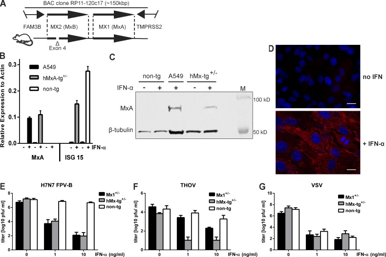
Cells from transgenic mice carrying the human MX locus exhibit IFN-dependent resistance to influenza A virus and THOV. (A) Schematic drawing showing the fragment of human chromosome 21 present in BAC clone Rp11-120c17 (top) and transgenic mice (bottom). Note that the MX2 gene is defective in our transgenic mice as a result of a deletion that includes coding exon 4. (B and C) MEFs prepared from transgenic (hMx-tg+/−) or nontransgenic (non-tg) littermates were treated with 10 ng per ml of IFN-α for 18 h (+) or were left untreated (–) before analysis of the cellular MxA content by RT-qPCR (B) or Western blot (C). IFN-treated human A549 cells served as positive control. ISG15 served as IFN treatment control in MEFs. Western blots were simultaneously probed with an antibody recognizing β-tubulin to demonstrate similar loading of the gel. RT-qPCR ΔCT values were calculated relative to actin gene expression from technical triplicates; SEM is shown. (D) MEFs from hMx-tg+/− mice were treated with 10 ng per ml of IFN-α for 18 h before visualization of MxA expression (red) by indirect immunofluorescence. Untreated cells served as controls. Nuclei were stained with DAPI (blue). Bars, 10 µm. (E) MEFs from hMx-tg+/− and non-tg littermates were treated for 18 h with plain medium (0) or medium containing 1 or 10 ng per ml of IFN-α before infection (MOI, 0.01) with H7N7 avian influenza A virus strain FPV-B. Culture supernatants were harvested at 24 h after infection and virus titers were determined. Cells from mouse embryos carrying one functional allele of the endogenous Mx1 gene (Mx1+/−) were used as controls. Mean values with standard error of means of three independent experiments are shown. (F and G) MEFs from hMx-tg+/− and non-tg littermates were treated for 18 h with plain medium (0) or medium containing 1 or 10 ng per ml of IFN-α before infection (MOI = 0.1) with THOV (F) or VSV (G). Culture supernatants were harvested at 24 h (VSV) or 72 h (THOV) after infection and virus titers were determined. Mx1+/− MEFs were used as controls. Mean values with SEM of three independent experiments are shown.

