Figure 7.
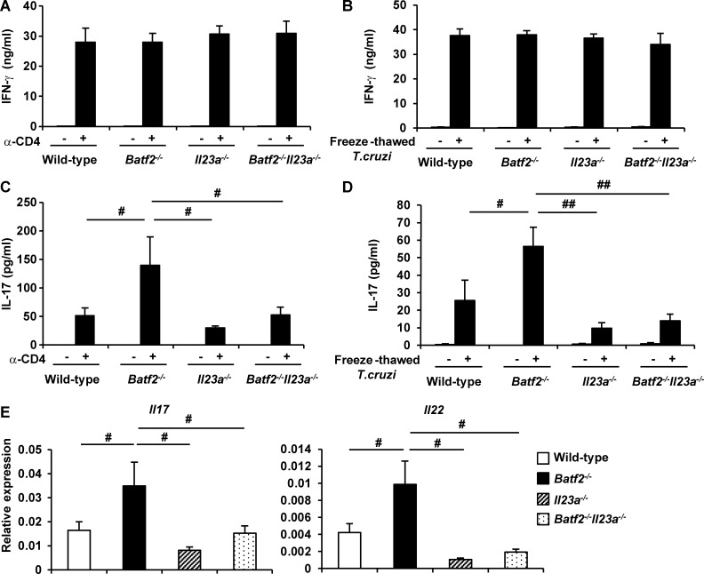
Reduced CD4+ T cell IL-17 production in T. cruzi–infected Batf2−/−Il23a−/− mice. (A–D) Wild-type (n = 18), Batf2−/− (n = 12), Il23a−/− (n = 9), and Batf2−/−Il23a−/− (n = 9) mice were infected with T. cruzi for 20 d. CD4+ T cells were isolated from the spleen of T. cruzi–infected mice and stimulated with anti-CD3 antibody (A and C) or freeze-thawed T. cruzi in the presence of antigen-presenting cells (B and D) for 24 h. The culture supernatants were analyzed for IFN-γ (A and B) and IL-17 (C and D) by ELISA. #, P < 0.045; ##, P < 0.006. (E) Expression of Il17a and Il22 in CD4+ T cells from the livers of T. cruzi–infected mice. #, P < 0.05. (A–E) Graphs show mean values ± SEM.

