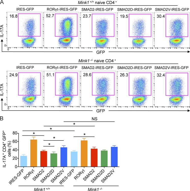
Figure 6.
MINK1 mediates Th17 cell differentiation by mediating SMAD2-α–helix 1 phosphorylation. (A) Sorted Mink1−/− and WT naive T cells were polarized under Th17 cell conditions as described in Fig. 3 A, except that the cells were first infected with the indicated retrovirus, and the cytokines were added 24 h later. The infection was repeated once at 48 h. 5 d later, the cells were analyzed for IL-17A+ cells after restimulation with PMA + ionomycin for 5 h. The infection efficiency was determined by GFP expression. The numbers in the graphs show the percentages of the gated populations. (B) Summary of retrovirus-infected Th17 cells as described in Fig. 6 A. Error bars show mean ± SD. *, P ≤ 0.05. n = 3 in each group; Student’s t test. Data are representative of two independent experiments. IRES, internal ribosomal entry site.

