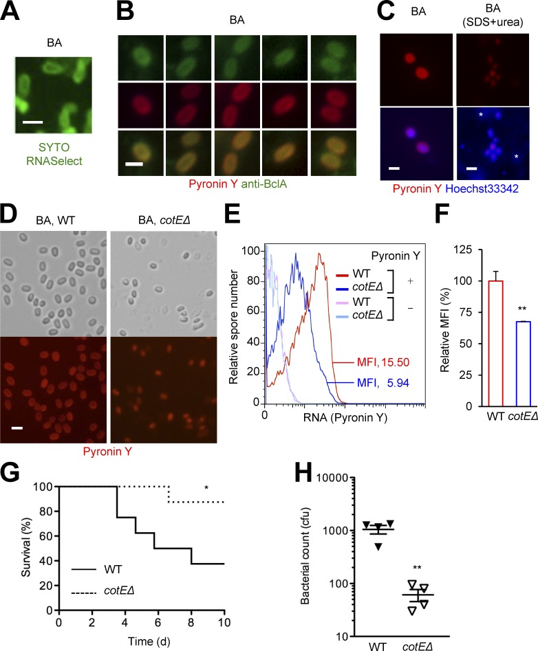
Figure 7.
BA spore RNA is mainly localized to the exosporium. (A–F) Spores of the indicated BA strains were stained for RNA (CYTO RNASelect and Pyronin Y), DNA (Hoechst 33342), and BclA (fluorescently labeled antibody) as indicated. Fluorescence signals from the labeling agents were analyzed by microscopy (A–D) or flow cytometry (E and F). Where indicated (C), BA spores were treated with SDS and urea before staining to eliminate the proteinaceous surface layers. *, nonspecific signals. Bar, 1 µm. MFI, mean fluorescence intensity. (G and H) Mice were infected with the indicated BA strains (107 and 2.5 × 10⁶ cfu per host; F and G, respectively) by footpad injection of spores. Host survival (n = 8 mice per group) was monitored for 10 d (G). Bacterial burdens in popliteal lymph nodes were determined 3 d after infection (H). Data are representative of three experiments (A, E, and F) or two experiments (B–D) with similar results, or from one experiment (G and H). *, P < 0.05; **, P < 0.01; Student’s t test (F and H) and Log-rank test (G).

