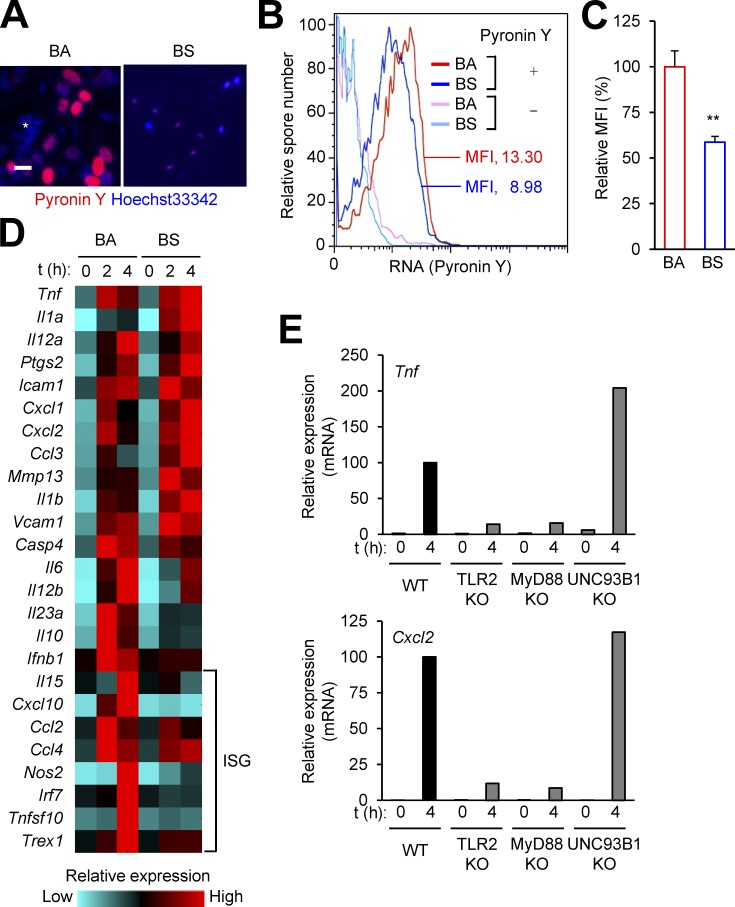
Figure 8.
BS spores do not depend on RNA for immunostimulation. (A–C) BA and BS spores were stained for RNA (Pyronin Y) and DNA (Hoechst 33342) as indicated. Fluorescence signals from the labeling agents were analyzed by microscopy (A) or flow cytometry (B and C). *, nonspecific signals. Bar, 1 µm. MFI, mean fluorescence intensity. **, P < 0.01; Student’s t test. (D and E) WT mouse macrophages (D) or macrophages from the indicated mice (E) were exposed to BA (D) or BS (D and E) spores at moi of 3. Relative mRNA amounts for spore-induced genes at the indicated time points after exposure were determined by qPCR, and presented in color-coded arbitrary units (D) or plotted on a linear scale (E). ISG, IFN-stimulated gene. Data are from one experiment (A, D, and E) or representative of three experiments (B and C) with similar results.

