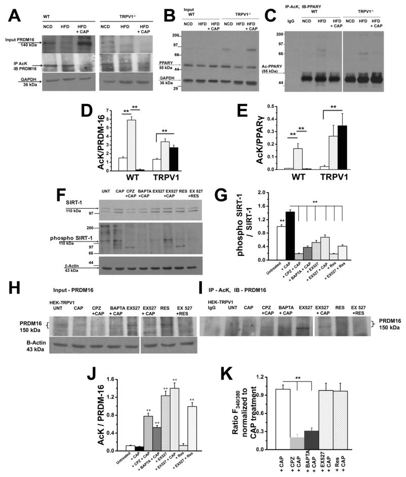
Figure 5. CAP increases deacetylation of PRDM-16 and PPARγ by activating SIRT-1.
A. Top panel shows western blot for PRDM-16 in 10% of total protein used for coimmunoprecipitation studies. Middle Panel shows acetylated PRDM-16 in BAT lysates of WT and TRPV1−/− mice-fed NCD or HFD (± CAP). Lysates were immunoprecipitated with acetylated lysine (Ac-K) antibody and immunoblotted for PRDM-16. Bottom panel represents GAPDH, loading control. B. Immunoblotting of 10% total protein used for coimmunoprecipitation with PPARγ and GAPDH. The proteins were resolved using 10% SDS-PAGE gel. C. Immunoprecipitation of samples using Ac-K antibody and immunoblotted with anti-PPARγ antibody. The samples were resolved using 7.5% SDS PAGE gel. D and E show densitometric ratios of acetylated PRDM-16 to total PRDM-16 and acetylated PPARγ to total PPARγ, respectively. F. Top panel shows the immunoblotting of lysates of TRPV1 expressing HEK 293 cells after treatment with CAP, CPZ + CAP, BAPTA-AM + CAP, EX527, EX527 + CAP, Res, and EX527 + Res with SIRT-1 antibody. Middle panel represents phosphorylated SIRT-1 on samples immunoprecipitated with SIRT-1 followed by immunoblotting with phosphoserine and phosphothreonine antibodies. Bottom panel shows β-actin loading control. G. Densitometric ratio of band intensities of phospho SIRT-1 to total SIRT-1. H. Immunoblot of TRPV1 expressing HEK 293 cells lysates after various treatments and probed with anti-PRDM16 and β-actin (loading control) antibodies. I. Immunoblot of acetylated PRDM-16 in TRPV1 expressing HEK 293 cells lysates after immunoprecipitation with AcK and immunoblotting with anti-PRDM-16 antibody. J. The densitometric band intensity ratios between acetylated PRDM-16 to total PRDM-16. (n = 6 independent experiments). K. Mean ratio ± S.E.M of Fura 2-AM fluorescence in TRPV1 expressing HEK 293 cells stimulated with CAP, CAP + CAP, BAPRA-AM + CAP, EX527 + CAP and Res + CAP (n = 88 – 122 cells). ** represent statistical significance for P<0.05.

