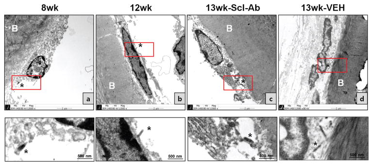
Figure 4. Electron microscopy analysis of the effect of Scl-Ab on LacZ (+) osteoblastic descendants on the periosteal surface of the calvaria.
X-gal stained calvarial bones were sectioned at 1μm thickness to visualize tissue morphology and to align X-gal staining with EM imaging. EM analysis was performed at 2 days (a: 8w) and 4 weeks (b: 12w) after the last 4-OHTam or after Scl-Ab (c) or vehicle (d) treatments twice a week. Each lower panel shows a magnified image of the red box in the upper panel. X-gal deposits in the cytoplasm of LacZ+ cells were visible by EM as black amorphous material (asterisk). Data are representative of experiments performed on sections from four mice for each group.

