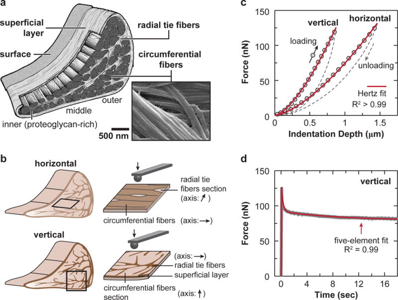
Fig. 1.

AFM-nanomechanical test set-up. (a) Schematic of the meniscus extracellular matrix (ECM) highlighting the four major structural units: surface, superficial layer, radial tie fibers and circumferential fibers, as well as the zonal variation. Scanning electron microscopy (SEM) image showing that two adjacent circumferential fibers are interconnected by fibrils spanning across them (white arrowhead). (b) Schematics of AFM-nanoindentation test on the horizontal and vertical sections, where the fiber axis orientation within each interior structural unit is shown. (c) Representative indentation force versus depth (F-D) curves measured on both horizontal and vertical sections of outer zone circumferential fibers, and corresponding Hertz model fit (tip radius R ≈ 5 μm, spring constant k ≈ 0.6 N/m, R2 > 0.99). (d) Representative relaxation curve of temporal force, F(t), versus time, measured on the vertical section of outer zone circumferential fibers, and corresponding five-element model fit (R2 = 0.99).
