Figure 7.
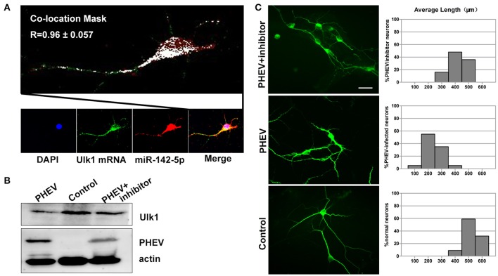
Enhanced Ulk1 by miR-142-5p inhibitors rescued axon elongation. (A) Co-localization assay of miR-142-5p (red) and Ulk1 mRNA (green) in primary cortical by Image J software. Pearson's R = 0.96 ± 0.057. (B) The PHEV-infected neurons were transfected with miR-142-5p inhibitors at a concentration of 200 nM. Forty-eight h after transfection, the cells were harvested for analysis of Western blot. (C) The PHEV infected neurons were transfected with miR-142-5p inhibitors at a concentration of 200 nM for 48 h. Antibody straining showed the infected neurons transfected with inhibitors did not have restricted axon elongation. Bar graph: quantification of neurons with different axon lengths. Data represent the means of n = 50 cells per condition counted in triplicate ± s.d. Scale bars are 50 μm in the image.

