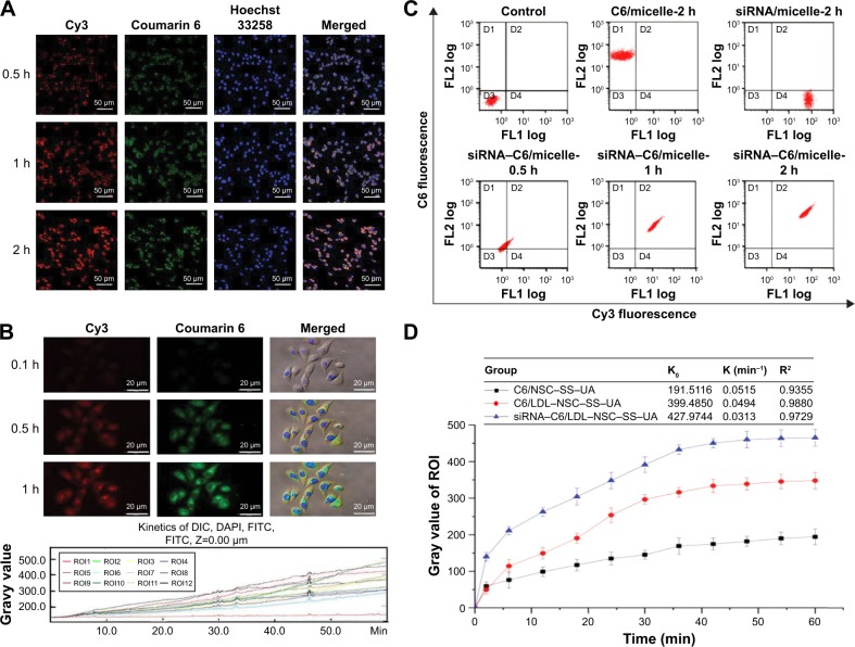
Figure 7.
Cellular uptake and distribution of the siRNA–PTX/LDL–NSC–SS–UA micelles in MCF-7/Taxol cells in vitro.
Notes: (A) Intracellular uptake and location of siRNA–PTX/LDL–NSC–SS–UA micelles in MCF-7/Taxol cells. (B) Dynamic uptake images of micelles and kinetics of fluorescence intensity. (C) Quantitative uptake of micelles in MCF-7/Taxol cells with two-color flow cytometry. (D) Micelle uptake kinetic parameters obtained by the change in fluorescence over time. Magnification 40×.
Abbreviations: Cy3, cyanine 3; C6, coumarin 6; siRNA–PTX/LDL–NSC–SS–UA, small interfering RNA–paclitaxel/low-density lipoprotein–N-succinyl chitosan–cystamine–urocanic acid; DIC, differential interference contrast; FITC, fluorescein isothiocyanate; DAPI, 4′,6-diamidino-2-phenylindole.

