Figure 6.
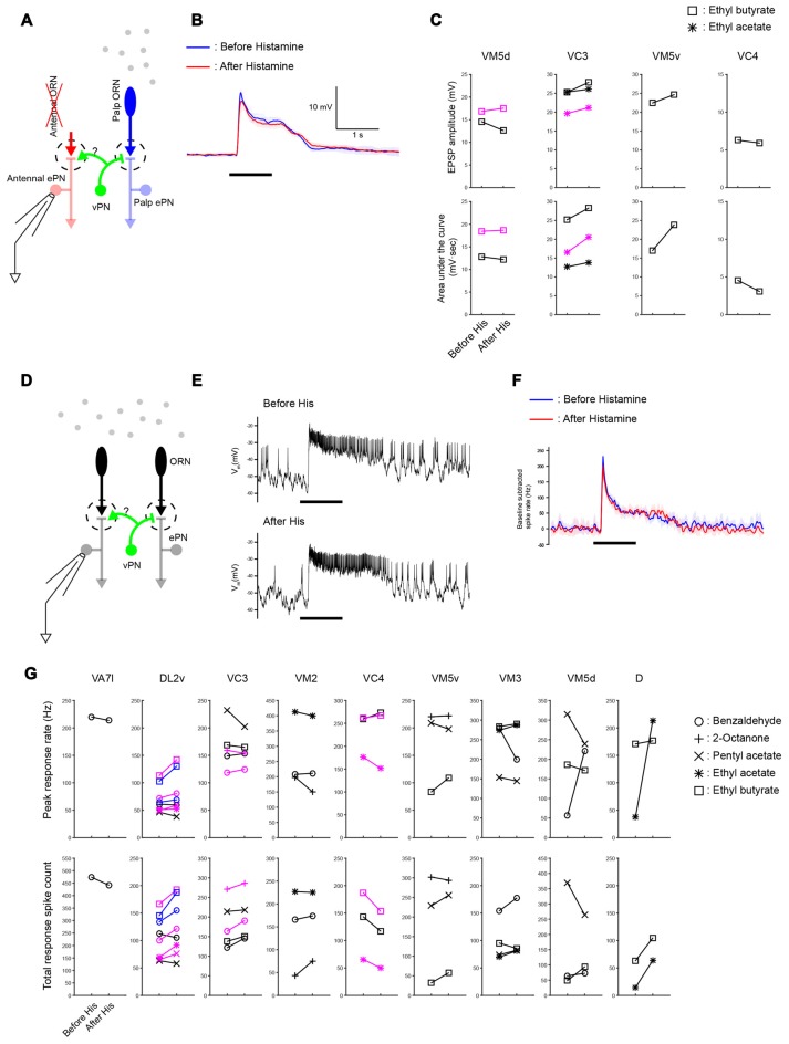
Widespread lateral excitation is not affected by suppressing vPNs. (A) A schematic of the antenna-less preparation. (B) Across-trial average of odor-elicited EPSPs from an example VM5d ePN (black plots in VM5d panel (C)) before and after suppressing vPNs expressing shunting His-Cl channels by adding histamine to the bath (mean ± SD) suggests that the vPN-ePN synapse does not contribute to lateral excitation. Odor stimulus in this example was a 1 s puff of 0.3% ethyl butyrate. (C) Results from similar experiments (six ePNs, seven odor-ePN pairs, p = 0.23 for the peak amplitude, p = 0.14 for the area under the curve, two-way ANOVA). Different ePNs in each glomerulus are indicated with different colors (black or magenta). (D) A schematic of the intact preparation. Both antennae and maxillary palps were stimulated with odors while ePNs were monitored with patch recordings. (E) Example voltage traces show odor-elicited responses in a VC3 ePN (black plots in VC3 panel (G)) before and after vPNs were suppressed by His-Cl channels. Odor stimulus was 1 s of 0.3% pentyl acetate. (F) Across-trial average of PSTHs from the same VC3 ePN shown in (E; mean ± SD). (G) Odor-elicited spiking responses in the palp ePNs (VA7l) and antennal ePNs (all others) were not reduced when vPNs were suppressed by His-Cl channels (13 ePNs, 32 odor-ePN pairs, p = 0.53 for the peak response rate, p = 0.13 for the total response spike count, two-way ANOVA, see “Materials and Methods” Section for response calculations). Different ePNs in each glomerulus are indicated with different colors (black, magenta or blue).

