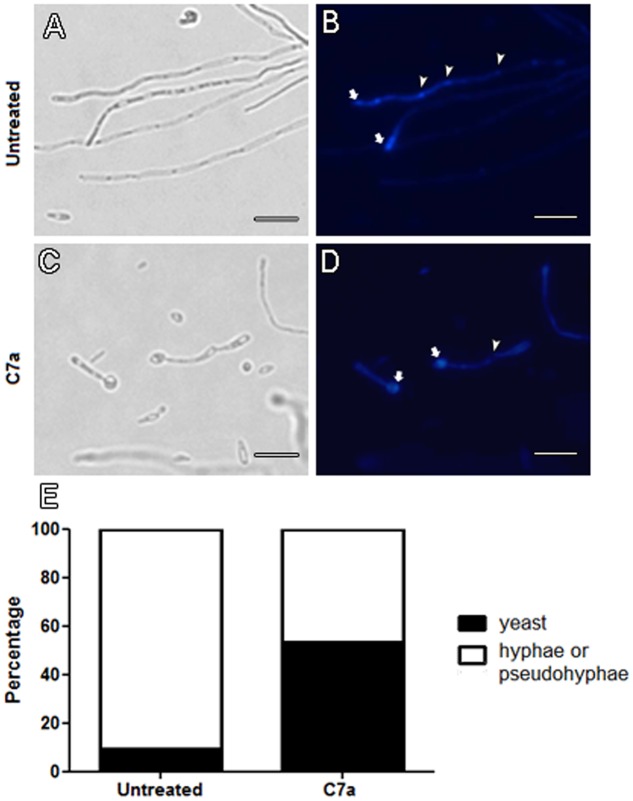
FIGURE 1.
Hyphae/pseudohyphae formation is impaired in C7a-treated cells. Yeast cells of Candida albicans ATCC 10231 were treated with 0.5 μg/mL of C7a for 24 h at 37°C in RPMI 1640 buffered with MOPS 0.16 M. Thereafter, untreated cells (A,B) and cells treated with C7a (C,D) were stained with Calcofluor White. Arrows in (B,D) show Calcofluor White staining at the end of hyphae and yeast-mother cell, respectively. Arrowheads in (B) show chitin-rich septa stained by Calcofluor White and in (D) absence of this structure. Yeasts and hyphae/psedohyphae were quantified as described in section “Material and Methods” (E). Bars = 20 micrometers.

