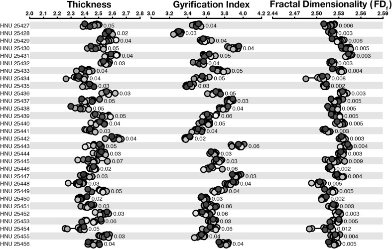
Fig. 1.
Dot plot for the structural estimates for each measure for the cortical ribbon, for the CCBD dataset. Participant labels are presented on the left, such that each row represents structural metrics for a single participant. Each dot within a measure (e.g., ‘Thickness’) represents a different scan volume. Within each row, markers in the same color denote measures taken from the same scan volume. Values beside each set of markers denote the mean deviation between estimates. (Color figure online)

