

Abstract
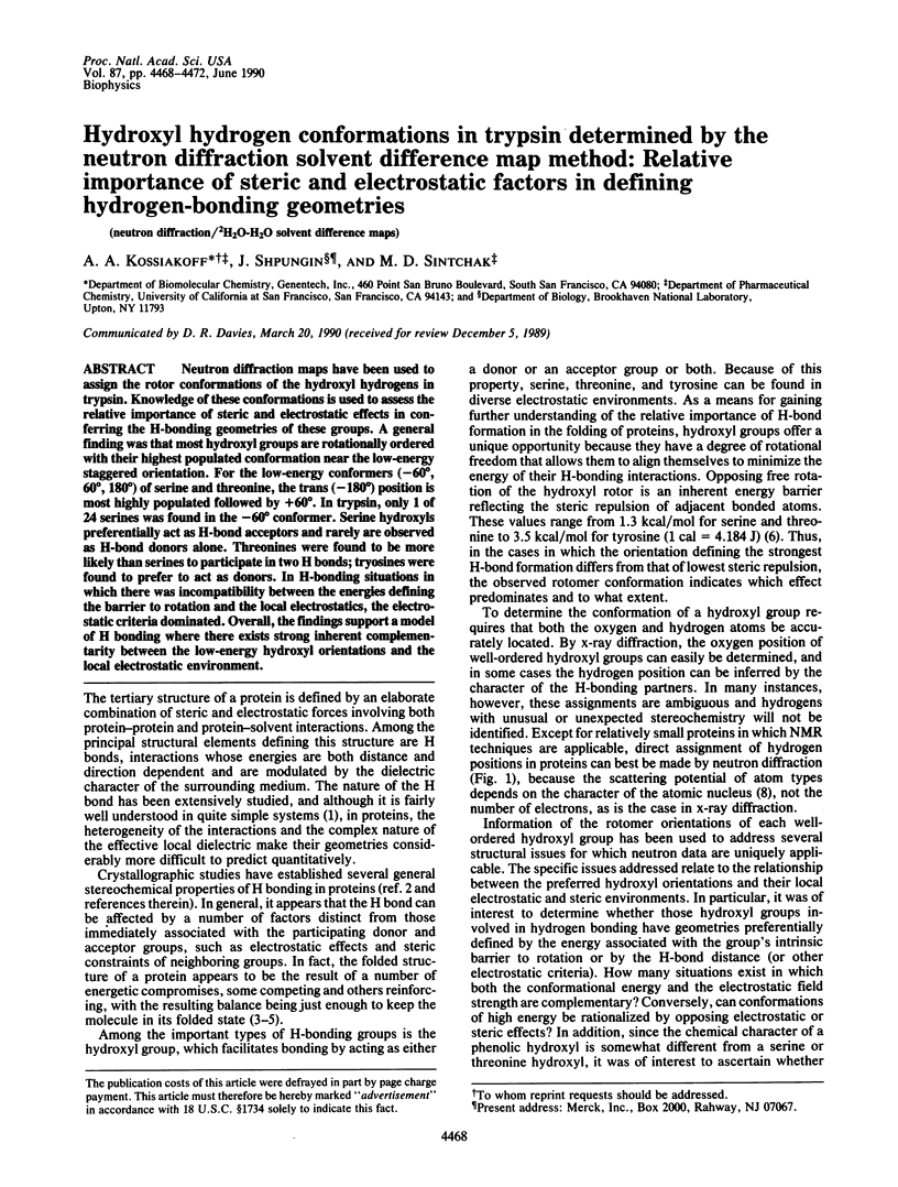
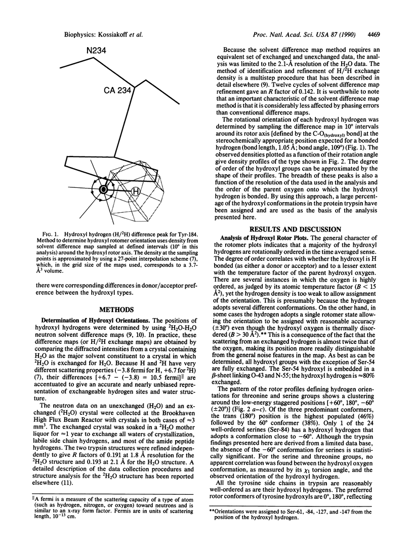
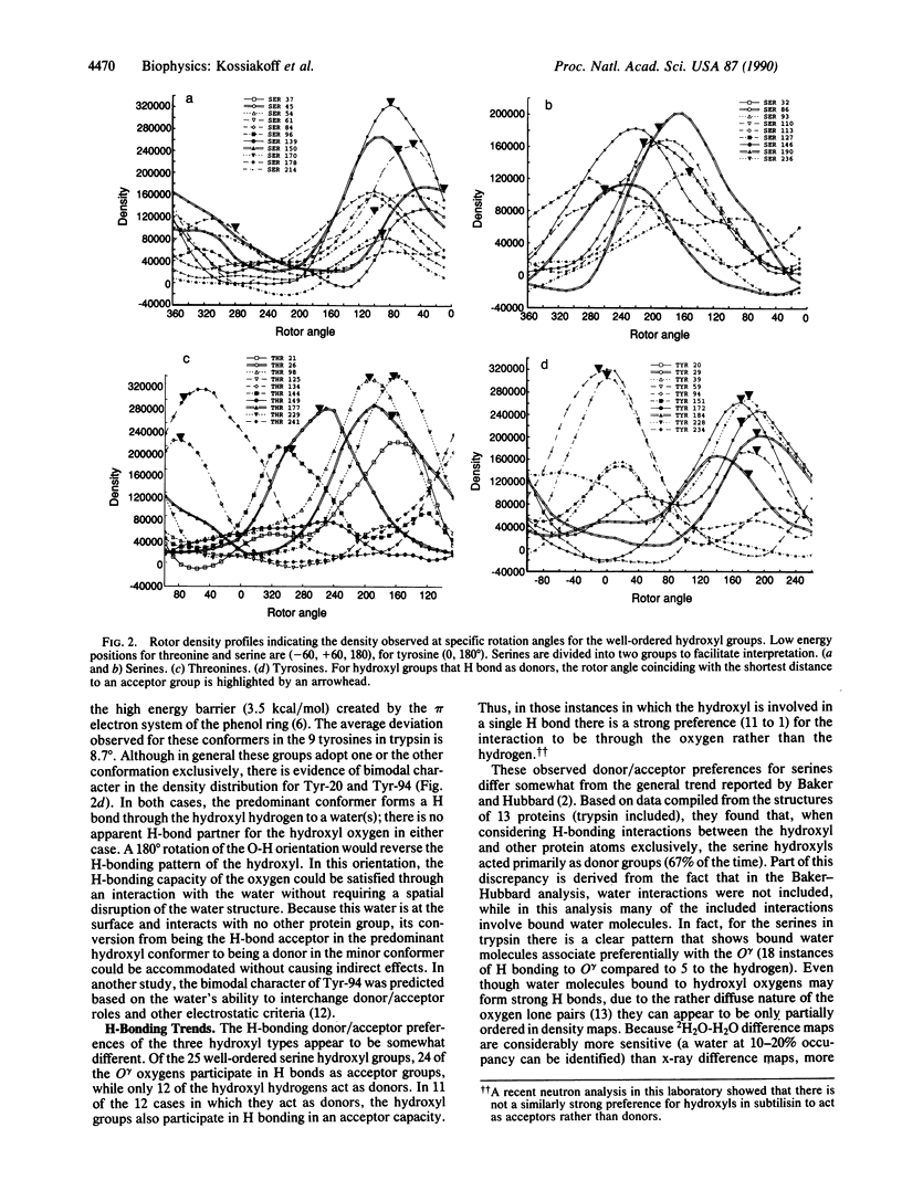
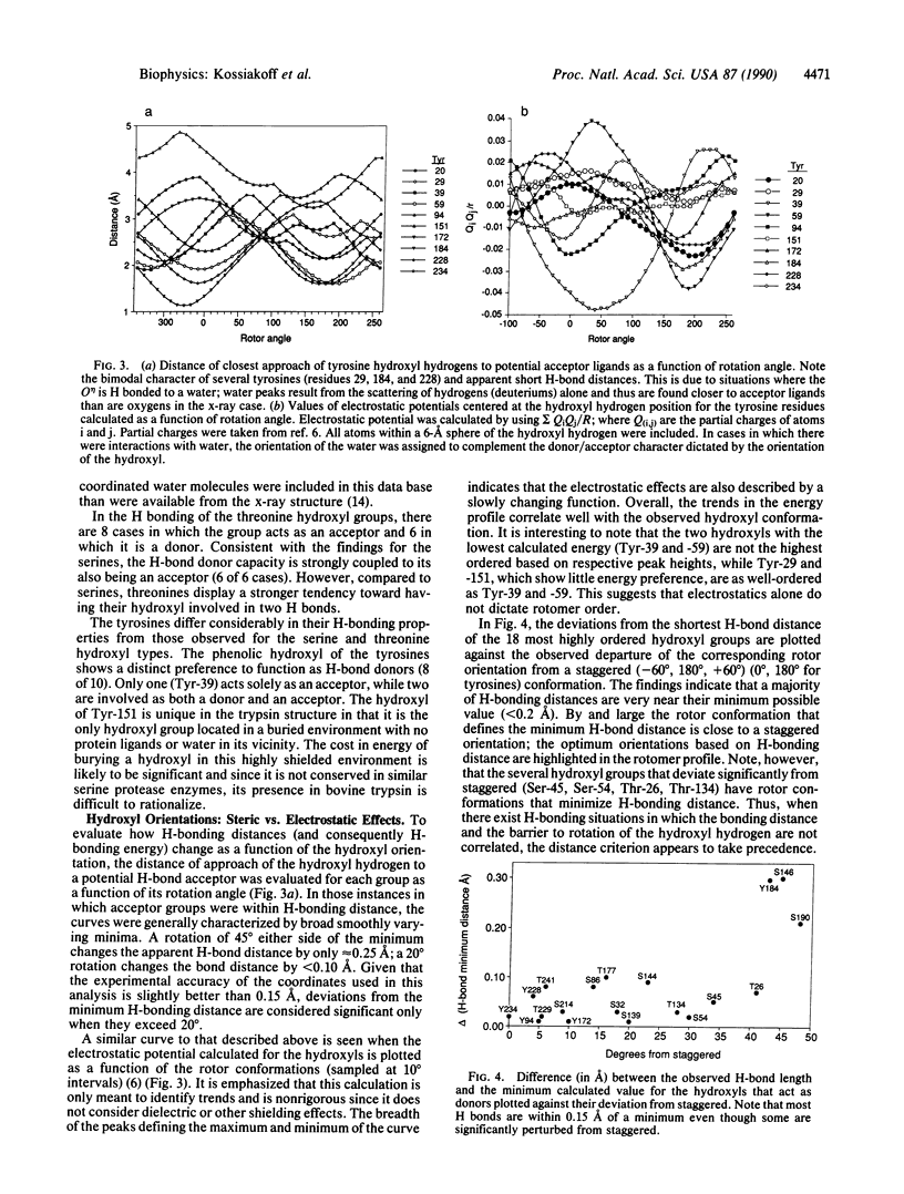
Neutron diffraction maps have been used to assign the rotor conformations of the hydroxyl hydrogens in trypsin. Knowledge of these conformations is used to assess the relative importance of steric and electrostatic effects in conferring the H-bonding geometries of these groups. A general finding was that most hydroxyl groups are rotationally ordered with their highest populated conformation near the low-energy staggered orientation. For the low-energy conformers (-60 degrees, 60 degrees, 180 degrees) of serine and threonine, the trans (-180 degrees) position is most highly populated followed by +60 degrees. In trypsin, only 1 of 24 serines was found in the -60 degrees conformer. Serine hydroxyls preferentially act as H-bond acceptors and rarely are observed as H-bond donors alone. Threonines were found to be more likely than serines to participate in two H bonds; tryosines were found to prefer to act as donors. In H-bonding situations in which there was incompatibility between the energies defining the barrier to rotation and the local electrostatics, the electrostatic criteria dominated. Overall, the findings support a model of H bonding where there exists strong inherent complementarity between the low-energy hydroxyl orientations and the local electrostatic environment.
Full text
PDF
Selected References
These references are in PubMed. This may not be the complete list of references from this article.
- Baker E. N., Hubbard R. E. Hydrogen bonding in globular proteins. Prog Biophys Mol Biol. 1984;44(2):97–179. doi: 10.1016/0079-6107(84)90007-5. [DOI] [PubMed] [Google Scholar]
- Bernstein F. C., Koetzle T. F., Williams G. J., Meyer E. F., Jr, Brice M. D., Rodgers J. R., Kennard O., Shimanouchi T., Tasumi M. The Protein Data Bank: a computer-based archival file for macromolecular structures. J Mol Biol. 1977 May 25;112(3):535–542. doi: 10.1016/s0022-2836(77)80200-3. [DOI] [PubMed] [Google Scholar]
- Brandts J. F., Hunt L. The thermodynamics of protein denaturation. 3. The denaturation of ribonuclease in water and in aqueous urea and aqueous ethanol mixtures. J Am Chem Soc. 1967 Sep 13;89(19):4826–4838. doi: 10.1021/ja00995a002. [DOI] [PubMed] [Google Scholar]
- Brünger A. T., Karplus M. Polar hydrogen positions in proteins: empirical energy placement and neutron diffraction comparison. Proteins. 1988;4(2):148–156. doi: 10.1002/prot.340040208. [DOI] [PubMed] [Google Scholar]
- Harrison R. W., Wlodawer A., Sjölin L. Analysis of solvent structure and hydrogen exchange in proteins on the basis of neutron diffraction data from deuterated and hydrogenous crystals. Acta Crystallogr A. 1988 May 1;44(Pt 3):309–320. doi: 10.1107/s010876738701242x. [DOI] [PubMed] [Google Scholar]
- KAUZMANN W. Some factors in the interpretation of protein denaturation. Adv Protein Chem. 1959;14:1–63. doi: 10.1016/s0065-3233(08)60608-7. [DOI] [PubMed] [Google Scholar]
- Kossiakoff A. A., Spencer S. A. Direct determination of the protonation states of aspartic acid-102 and histidine-57 in the tetrahedral intermediate of the serine proteases: neutron structure of trypsin. Biochemistry. 1981 Oct 27;20(22):6462–6474. doi: 10.1021/bi00525a027. [DOI] [PubMed] [Google Scholar]
- Meyer E., Cole G., Radhakrishnan R., Epp O. Structure of native porcine pancreatic elastase at 1.65 A resolutions. Acta Crystallogr B. 1988 Feb 1;44(Pt 1):26–38. doi: 10.1107/s0108768187007559. [DOI] [PubMed] [Google Scholar]
- Shpungin J., Kossiakoff A. A. A method of solvent structure analysis for proteins using D2O-H2O neutron difference maps. Methods Enzymol. 1986;127:329–342. doi: 10.1016/0076-6879(86)27027-5. [DOI] [PubMed] [Google Scholar]
- Tanford C. Protein denaturation. Adv Protein Chem. 1968;23:121–282. doi: 10.1016/s0065-3233(08)60401-5. [DOI] [PubMed] [Google Scholar]