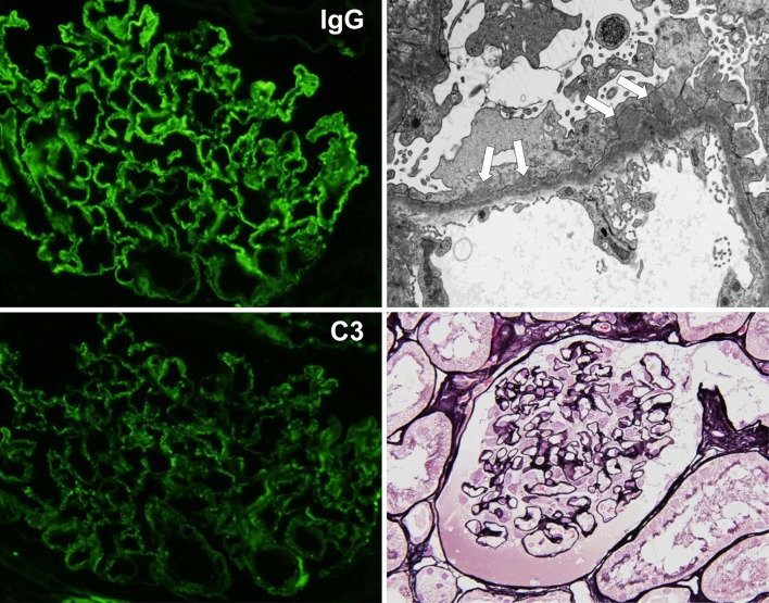
Fig. 1.
Findings of renal biopsy (first biopsy). The biopsy specimen showed 23 glomeruli, and 5 of them were deteriorated. Periodic acid silver-methenamine (PAM) staining of the non-deteriorated glomeruli revealed the partial formation of stippling and spike at the capillary. The immunofluorescence (IF) method revealed positive granular patterns of IgG and C3 along the glomerular capillary wall, and electron microscopy showed subepithelial electron-dense deposits. There were no findings suggestive of diabetic nephropathy, such as mesangial matrix accumulation, nodular glomerulosclerosis, and arteriolar hyalinosis. Membranous nephropathy (stage I–II) was diagnosed on the basis of these findings

