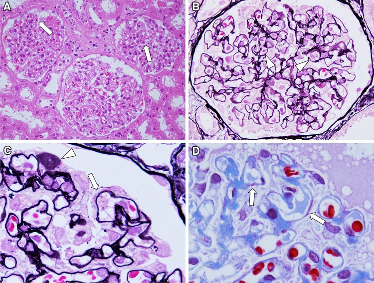
Fig. 1.
Light microscopy findings. Renal biopsy samples included 44 glomeruli and 1 glomerulus underwent obsolescence. Remaining all glomeruli showed diffuse but irregular thickening glomerular capillary walls (arrow in a). No apparent mesangial hypercellularity was noted in glomeruli (HE stain, ×400). In glomeruli, infiltrating cells in capillary lumens were seen (arrowhead in b) (PAM stain, ×600). High magnification of glomeruli showed focal and segmental formation of spike (arrow in c) and stippling (arrowhead in c) in glomerular basement membrane (PAM stain, ×1,000). Focal and segmental subepithelial side of deposits was also noted on glomerular capillary walls (arrow in d) (Masson stain, ×1,000)

