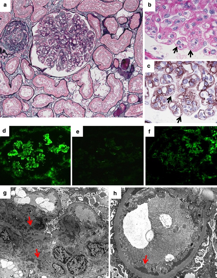
Fig. 2.
Second renal biopsy. a Glomerular capillary endotheliosis. Capillary lumina were narrowed or obstructed by marked mesangial and endothelial cell swelling and hypertrophy. Global sclerosis of glomeruli and 20 % interstitial fibrosis and tubular atrophy with foam cells (periodic acid silver methenamine–Masson trichrome, ×200). b Endothelial cell swelling indicated by black arrows (periodic acid-Schiff, ×1000). c Doubling of the basement membrane in glomeruli indicated by black arrows (periodic acid silver methenamine, ×1000). d Diffuse mesangial and partial capillary deposit of IgM (×400). e C3c deposit was minimal (×400). f Diffuse mesangial and partial capillary deposit of fibrinogen (×400). g Electron-dense deposit in mesangial region (red arrows, ×2500). h Electron-dense deposit in subendothelial region (red arrows, ×7000)

