
Figure 3.
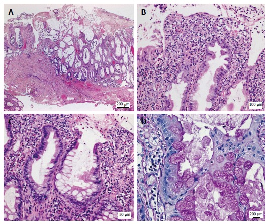
Histologic appearance of the colonic mucosa. A: Erosive lesions covered by a thick pseudomembranous layer; damage of the surface epithelium and architectural disarray of the mucosa showing prominent cystic dilatation of crypts; B: Foci of mildly serrated epithelium; C, D: Abundant hyperplastic goblet cells associated with a strikingly increased accumulation of intracellular and extracellular mucus. A-C: Hematoxylin and eosin; D: Periodic acid-Schiff reaction.