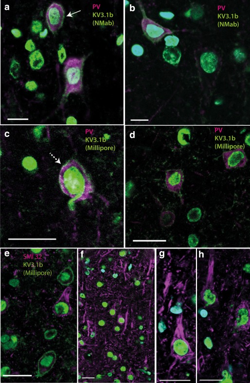
Figure 2.

Kv3.1b expression in rat motor cortex. Transverse sections of rat motor cortex labeled with Kv3.1b (green) antibodies and cell markers (magenta) as well as nuclear marker DAPI (blue). (a, b). The intensity of the membrane labeling of the neuron in a (solid arrow), which expressed Kv3.1b (NeuroMab, green) antibody and rabbit anti‐parvalbumin (magenta), is marked as “2A” in Figure 3b. (c, d) Kv3.1b (Millipore, green) antibody and mouse anti‐parvalbumin (magenta). (e–h) Examples of labeling for Kv3.1b (Millipore, green) antibody and mouse anti‐SMI32 (magenta). Note that neurons positively stained for SMI32 have pyramidal morphology but no clear Kv3.1b staining in the membrane. Scale bars: 20 µm [Color figure can be viewed at wileyonlinelibrary.com]
