
Figure 3.
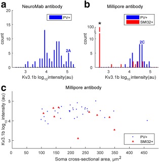
Quantification of Kv3.1b labeling in rat motor cortex. The total intensity of Kv3.1b labeling in the membrane of each neuron was quantified using MetaMorph Offline software and expressed in arbitrary units (au). It was converted to a log10 scale. (a) Distribution of total intensities for staining with the NeuroMab antibody in 67 parvalbumin‐positive putative interneurons (PV+). The marker 2A indicates the intensity value for the PV + labeled neuron shown in Figure 2a. (b) The distribution of Kv3.1b staining intensity is shown for 32 neurons which stained with the Millipore antibody and were also parvalbumin‐positive (PV+) (blue bars). The marker 2C indicates intensity value for the PV + labeled neuron shown in Figure 2c. The red bars indicate Kv3.1b staining intensity for nine neurons which were also co‐labeled with the pyramidal cell marker SMI32. The other 91 neurons analyzed which expressed the SMI32 marker showed no Kv3.1v staining, and are indicated by the tall red column on the far left (*). (c) The intensity of Kv3.1b staining with the Millipore antibody has been plotted against the soma cross‐sectional area of each labeled cell for PV‐positive (blue circles) and SMI32‐positive neurons (red triangles). Only a few SMI32‐positive pyramidal cells expressed Kv3.1b [Color figure can be viewed at wileyonlinelibrary.com]