Figure 4.

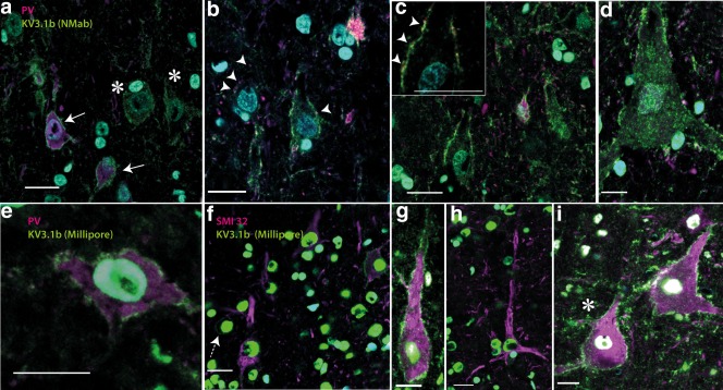
Kv3.1b expression in macaque motor cortex. Transverse sections of macaque motor cortex labeled with Kv3.1b (green) antibodies and cell markers (PV and SMI32; magenta and DAPI in blue). (a–d) Kv3.1b (NeuroMab, green) antibody and anti‐parvalbumin (magenta). Parvalbumin‐positive neurons (solid arrows bottom left in (a) showed Kv3.1b membrane staining, but in addition, large, parvalbumin‐negative cells, with pyramidal cell morphology also strongly expressed Kv3.1b (asterisks). Further examples are shown in (b–d). High magnification revealed that pyramidal cells had numerous parvalbumin‐positive (magenta) synaptic boutons on their membranes (putative interneuron axon terminals; arrowheads in (b) and inset in (c), synaptic contact points shown as arrowheads), which contrasted with the Kv3.1b labeling present on these cells all along the membrane. (e–i) Show Kv3.1b labeling in neurons using the Millipore antibody (green) and either parvalbumin (e) or SMI32 antibody (f–i) (both magenta). The Millipore antibody stained the cell membrane in parvalbumin‐positive cells (e) but also labeled the membrane of large SMI32‐postitive pyramidal cells (g and i). (f) Shows four SMI32 positive pyramidal neurons, one of which (top right) is also positive for Kv3.1b. A small round SMI32‐negative neuron with clear Kv3.1b labeling is also present in this image (dashed arrow). (h) Shows another SMI32‐positive pyramidal cell that did not express Kv3.1b. Scale bars: 20 µm [Color figure can be viewed at wileyonlinelibrary.com]
