
Figure 2.
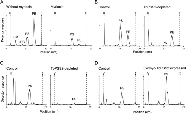
Lipid analysis of TbPSS2 parasites after [3H]serine labeling. Procyclic form trypanosomes or digitonin extracts were labeled with [3H]serine for 4 h and lipids were extracted, separated by one‐dimensional thin layer chromatography and detected using a radioisotope detector.
A. [3H]Serine labeling of phospholipids in parasites before (left panel) and after 30 min of myriocin treatment (2.5 µM; right panel).
B, C. [3H]Serine labeling of whole cells (panel B) and digitonin extracts (panel C) before and after RNAi‐mediated depletion of TbPSS2 for 6 days.
D. [3H]Serine labeling of control and 3xc‐myc‐TbPSS2 overexpressing procyclic forms after 2 days of tetracycline induction. The scans are representative of at least three independent experiments.