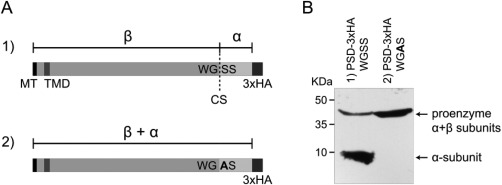
Figure 3.

Processing of TbPSD.
A. Predicted processing of wild‐type (1) and mutated (2) TbPSD into α‐ and β‐subunits. MT, mitochondrial targeting signal; TMD, transmembrane domain; CS, cleavage site; WGSS/WGAS, amino acid motifs at predicted cleavage site; 3xHA, C‐terminal attachment of 3xHA tag.
B. SDS‐PAGE/immunoblot showing two bands representing the proenzyme (43.2 kDa) and the cleaved α‐subunit (7.4 kDa) (left lane), and a single band representing the uncleaved proenzyme after mutation of the recognition site (right lane).
