
Figure 4.
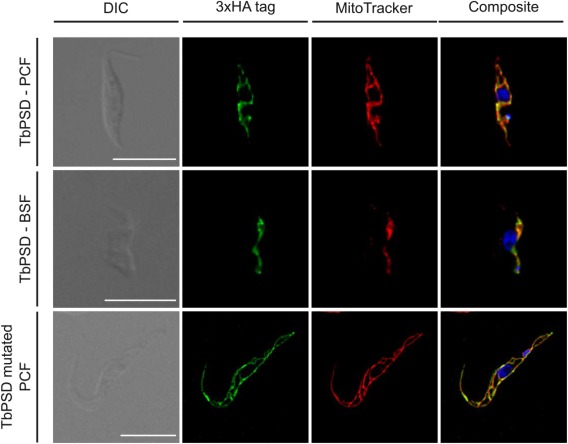
Localization of TbPSD by immunofluorescence microscopy. Hemagglutinin (HA)‐tagged wild‐type (top and middle rows) and mutated forms (bottom row) of TbPSD were expressed in T. brucei procyclic (PCF; top and bottom rows) and bloodstream forms (BSF; middle row). Subcellular localization of TbPSD was analyzed by immunofluorescence microscopy using anti‐HA antibody and the mitochondrial dye MitoTracker. DNA (in blue) was stained with 4′,6′‐diamidino‐2‐phenylindole. Scale bars indicate 10 µm.