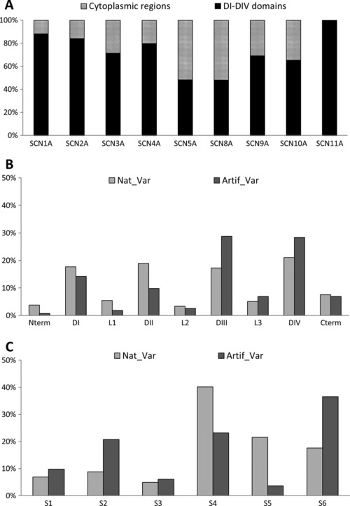
Figure 3.

Distribution of the variants according to the topological domains: A: DI–DIV domains versus cytoplasmic regions. Distribution of the variants with electrophysiological defects: the natural variants (Nat_Var) versus artificial variants (Artif_Var) according to (B) the domains or to (C) the segments
