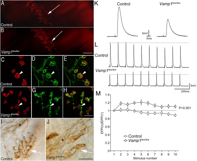
Figure 2.

Synaptic defects at the neuromuscular junctions in Vamp1 lew/lew mice. (A, B) Low‐power images of the whole‐mount diaphragm muscles (P14) labeled by Texas Red—conjugated α‐bungarotoxin. The endplate band (arrow) is similarly localized along the central regions of the muscle in both control (A) and Vamp1 lew/lew mice (B). (C–H) High‐power confocal images of individual neuromuscular synapses in triangularis sterni muscles, labeled by Texas Red–conjugated α‐bungarotoxin (arrowheads in C and F) and antineurofilament NF150 and antisynaptotagmin2 antibodies (arrowheads in D and G point to the nerve terminals). Merged images are shown in E and H, for control and Vamp1 lew/lew mice, respectively. (I, J) Individual neuromuscular synapses (arrows) in triangularis sterni muscles labeled by antisyntaxin1 antibodies. The synapses are markedly smaller in Vamp1 lew/lew mice compared with the control. Asterisks indicate nerve bundles. (K) An example of endplate potentials (EPPs) recorded in the diaphragm muscle in control and Vamp1 lew/lew mice. (L) EPP traces responding to a low‐frequency, repetitive nerve stimulation (10Hz). (M) Quantitative measurement of the ratios of EPP amplitudes: EPP(n) to the first EPP amplitude, (EPP1). A low‐frequency, repetitive stimulation (10Hz) led to a run‐down of EPPs in control, but synaptic facilitation in (Vamp1 lew/lew) mice.
