
Figure 3.
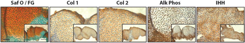
Limb bud mesenchymal micromass. Embryonic chick limb bud micromass cultured for 21 days and stained with Safranin O/Fast green (Saf O/FG), and immunostained for collagen type I (Col 1), collagen type II (Col 2), alkaline phosphatase (Alk Phos), and Indian hedgehog (IHH). Bar =50 μm, Inset Bar 100 μm. (A color version of this figure is available in the online journal.)