
Figure 4.
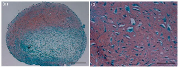
MSC pellet culture. Human bone marrow-derived MSCs were pelleted and cultured in TGF-β1 containing chondrogenic medium for 21 days. Samples were processed for histology and stained with Safranin O/Fast green. Red staining indicates matrix sulfated glycosaminoglycan deposition. Bar =250 μm in a and 50 μm in b. (A color version of this figure is available in the online journal.)