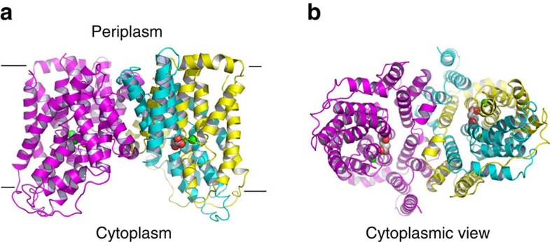
Figure 1. Structure of the succinate-bound VcINDY.
(a) Structure of dimeric VcINDY as viewed from the membrane bilayer. VcINDY is shown in ribbon rendition, the N (18–231) and C (232–462) domains in one protomer are coloured cyan and yellow, respectively, whereas the other protomer is coloured magenta. Na+ ions (green) and succinate are drawn as spheres. (b) The cytoplasmic view of the VcINDY structure, highlighting the solvent-accessible succinate and buried Na+ ions.

