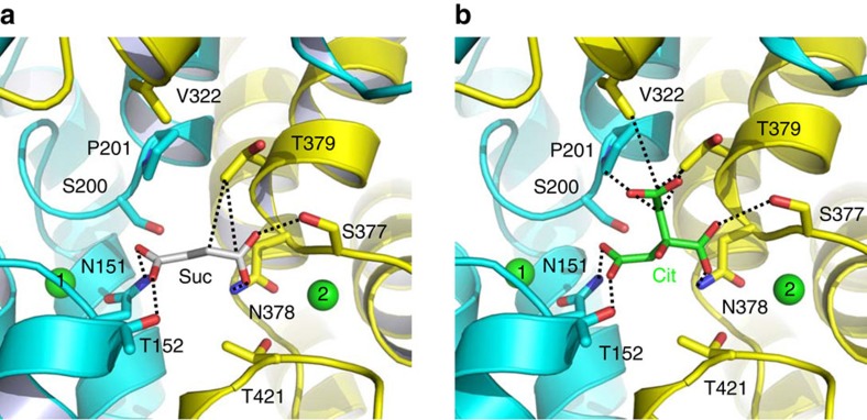
Figure 5. Close-up views of the succinate- and citrate-binding sites in VcINDY.
(a) Structure of the succinate-binding site. (b) Detailed view of the citrate-binding site. Succinate (grey), citrate (green) and relevant amino acids are drawn as stick models, whereas the Na+ ions are shown as green spheres. Dashed lines highlight the interactions between VcINDY and succinate or citrate.

