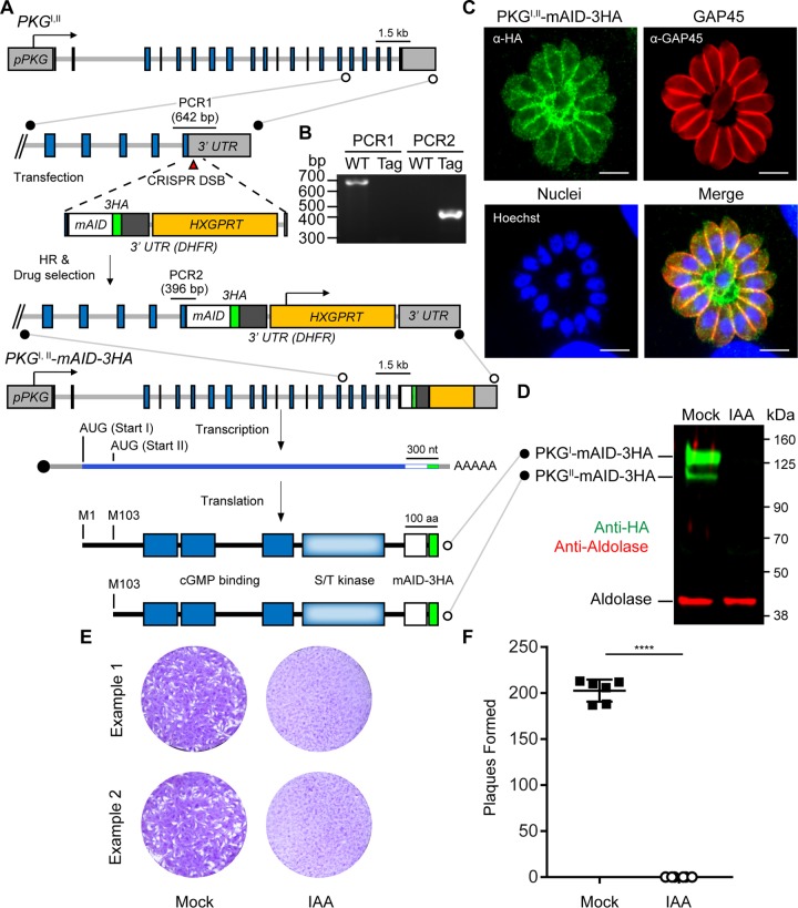
FIG 2 .
Fusion of mAID to PKGI,II in TIR1 parasites allows the simultaneous depletion of PKGI and PKGII isoforms. (A) Strategy for tagging of PKGI,II with mAID in TIR1-3FLAG parasites and depiction of the two protein isoforms produced from the PKGI,II-mAID-3HA transcript. (B) DNA electrophoretogram of diagnostic PCRs from genomic DNA showing 3′ integration of mAID-3HA into PKGI,II. The genomic loci acting as templates for PCR1 and PCR2 amplicons are shown in panel A. Lanes: WT (wild type), TIR1-3FLAG parent; Tag, PKGI,II-mAID-3HA parasites. (C) Coexpression of PKGI-mAID-3HA and PKGII-mAID-3HA (both green) in PKGI,II-mAID-3HA parasites determined by IF microscopy. GAP45 (red) is a marker for the parasite plasma membrane. Scale bars = 5 µm. (D) Western blot assay of lysed PKGI,II-mAID-3HA parasites probed with antibodies recognizing HA and aldolase. Parasites were treated with 500 µM IAA or the vehicle (EtOH) for 4 h prior to lysis. (E, F) Plaque formation by PKGI,II-mAID-3HA parasites treated with 500 µM IAA or the vehicle (EtOH) for 8 days (E) and mean number of plaques formed per well (mock, n = 6; IAA, n = 6) ± the standard deviation (F). ****, P < 0.0001 (unpaired two-tailed Student t test). Panels D to F each show data from a single example from three experiments with the same outcome.

