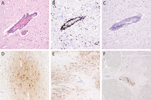
Figure 2.

Immunohistochemistry depicting inflammatory reaction and eastern equine encephalitis virus (EEEV) in the central nervous system are depicted. Perivascular and parenchymal chronic inflammatory infiltrates in a section of frontal cortex (A), consisting predominantly of CD3+ T lymphocytes (B) with no CD79a+ B lymphocytes (C). The EEEV is shown in neuronal cytoplasm in cortex (D), thalamus (E), and anterior horn cells in the thoracic spinal cord (F). Original magnification, ×40 (E and F), ×100 (D), and ×200 (A–C); hematoxylin and eosin stain (A); immunostains CD3 (B), CD79 (C), and EEEV (D–F).
