
Figure 2. Provision of exogenous human Flt3L and IL-15 enhances human NK cell reconstitution in humanized mice.
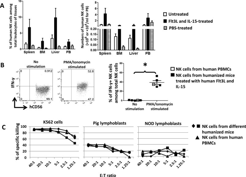
Humanized mice were generated by injection of human fetal liver-derived CD34+ cells to irradiated NSG mice. 14 weeks later, when mice were fully reconstituted by human cells, plasmid encoding human Flt3L was administered by hydrodynamic injection to these mice followed by injection of human IL-15. (A) Humanized mice without treatment (n=3) or treated with PBS (n=4) were used as controls for humanized mice treated with human Flt3L and IL-15 (n=4). Human NK cells in tissues were quantified. BM: bone marrow. PB: peripheral blood. Error bars represent SEM. *, p<0.05 with ANOVA, compared with PBS-treated group. (B) Splenocytes from humanized mice treated with human Flt3L and IL-15 were stimulated with PMA/Ionomycin for 5 hours in the presence of Brefeldin A followed by intracellular staining to determine the production of IFN-γ by human NK cells. Error bars represent SEM. *, p<0.05 with a Student’s t-test. NK cells from human PBMCs served as a control and were not included in the statistical analysis. (C) Human splenic NK cells from the spleen of humanized mice were isolated and their cytotoxic responses to pig lymphoblasts, K562 cells and NOD mice lymphoblasts were determined. Effector:target (E:T) ratio is shown on the x-axis. NK cells from human PBMCs served as a control. SP: spleen. BM: bone marrow. PB: peripheral blood.