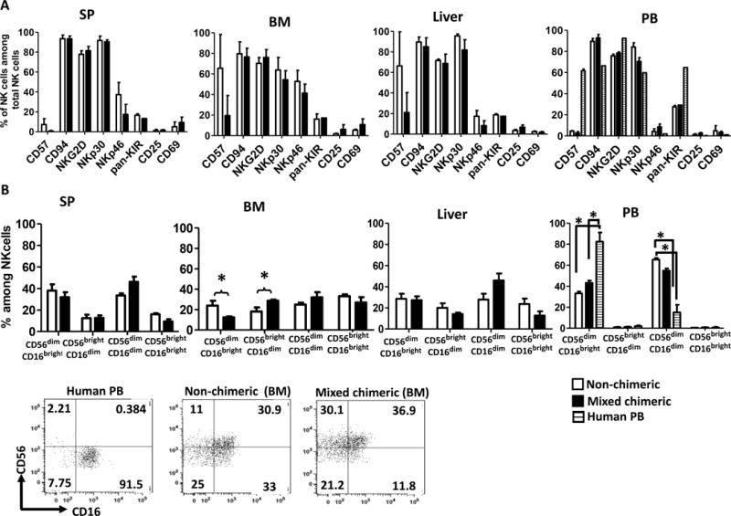
Figure 6. Phenotypic characterization of NK cells in mixed chimeras.

Pig/human mixed xenogeneic chimeras and non-chimeric mice were given hydrodynamic injection of plasmid encoding human Flt3L followed by injection of human IL-15 for 2 weeks. Mice were euthanized at the end of the 2-week Flt3L and IL-15 treatment and phenotype of human NK cells in various tissues was determined. (A) Phenotype of human NK cells in various tissues from pig/human mixed chimeric mice not showing global hyporesponsiveness (n=4) and non-chimeric mice (n=3). (B) Human NK cell subsets in various tissues identified by CD56 and CD16 expression. Representative flow cytometric dot plots from one experiment are shown to demonstrate the designation of NK cell subsets. Human peripheral blood NK cells (left) and bone marrow NK cells of non-chimeric and mixed chimeric mice are shown. Error bars represent SEM. *, p<0.05, Student’s t-test, comparison as specified in the figure. SP: spleen. BM: bone marrow. PB: peripheral blood.
