Figure 4.
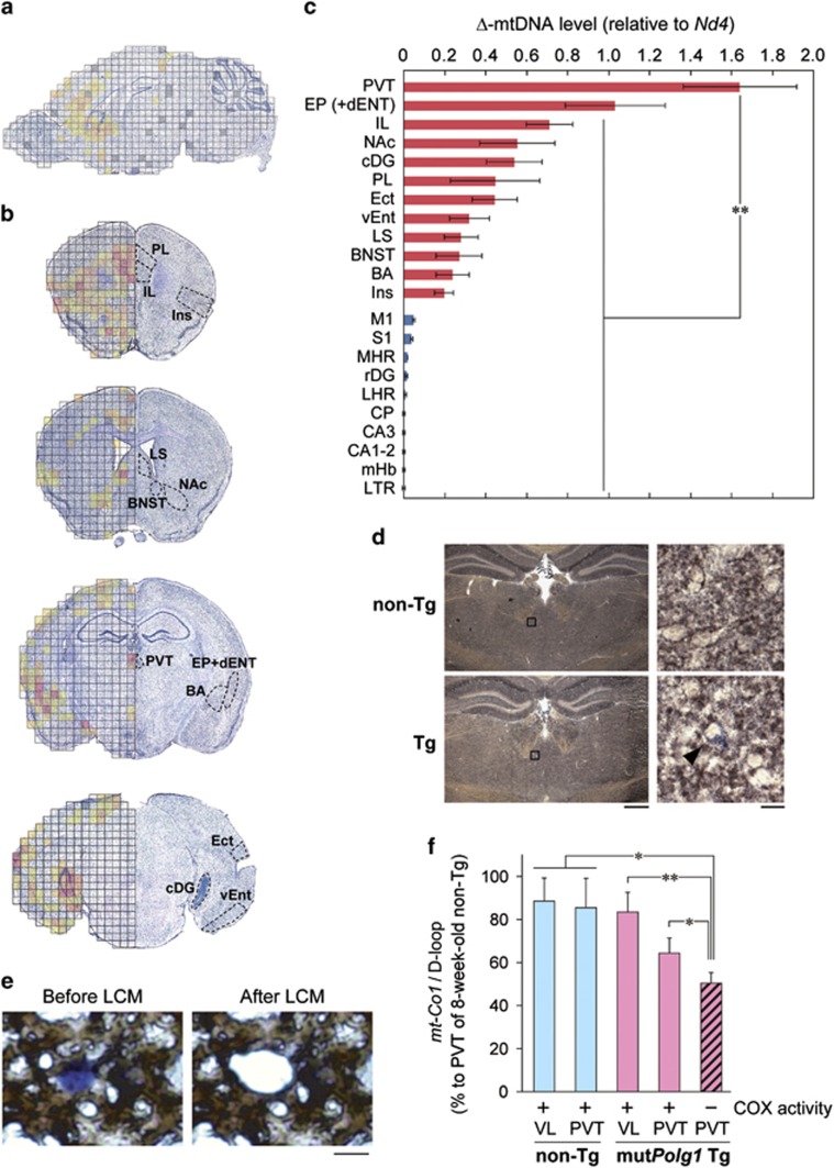
Accumulation of mtDNA deletions in the PVT of Polg1 mutant mice. (a) Comprehensive mapping of Δ-mtDNA of a Polg1 mutant mouse. Δ-mtDNA levels are shown in the following colors: red (high), orange, light orange, gold, yellow (low) and no color (unquantifiable). Gray, not determined. (b) Mapping of Δ-mtDNA levels on four coronal sections of a Polg1 mutant mouse. Regions with higher levels of Δ-mtDNA are marked. (c) Quantitative Δ-mtDNA levels in regions of interest, where higher (red) and lower (blue) levels of Δ-mtDNA are suggested. n=5 (individual mice) per region. **P<0.01. (d) Activity staining of SDH (blue) and COX (brown) of coronal sections at the level of PVT. The right images are magnified views of the boxed regions (left). A cell without COX activity (COX-negative cell) in the PVT is indicated by an arrowhead. Scale bars, 0.5 mm (left) and 10 μm (right). (e) Laser capture microdissection (LCM) of COX-negative cells after COX activity staining. Scale bar, 10 μm. (f) Quantification of Δ-mtDNA levels in LCMed COX-negative and -positive cells. The copy number ratio of mt-Co1/D-loop in LCMed cells inversely reflects the Δ-mtDNA level, and the ratio for the PVT of an 8-week-old wild-type mouse, assumed to have no deletion, is set to 100%. For example, the mt-Co1/D-loop ratio of 60% means that 40% of mtDNA molecules are Δ-mtDNAs. Data are expressed as means±s.e.m. of three mice. *P<0.05, **P<0.01. BA, basal amygdala; BNST, bed nucleus of the stria terminalis; CA, corpus ammonium; cDG, dentate gyrus, caudal part; CP, caudate putamen; dENT, entorhinal area, deep layer; Ect, ectorhinal area; EP, endopiriform nucleus; IL, infralimbic cortex; Ins, insular cortex; LHR, lateral hypothalamic region; LS, lateral septum; LTR, lateral thalamic region; M1, primary motor cortex; mHb, medial habenula; MHR, midline hypothalamic region; Δ-mtDNA, deleted mtDNA; NAc, nucleus accumbens; PL, prelimbic cortex; PVT, paraventricular thalamic nucleus; rDG, dentate gyrus, rostral part; S1, primary somatosensory cortex; vENT, entorhinal area, ventral part; VL, ventral lateral nucleus of the thalamus (See Supplementary Table 3).

