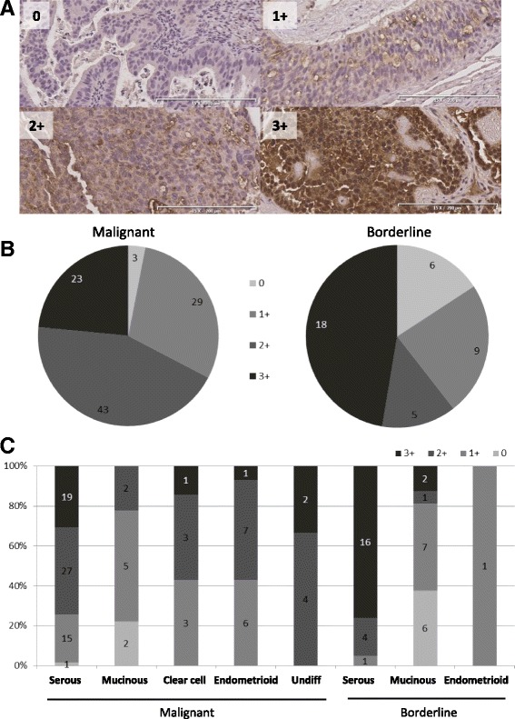
Fig. 2.

Illustrative images of the staining intensities and the distribution of the different intensities among the samples. a) Representative images demonstrating the different staining intensities Upper left: no staining = 0, serous (highly differentiated stage I), Upper right: 1 = weak staining (endometrioid poorly differentiated, stage II). Lower left: 2 = moderate staining (endometrioid poorly differentiated stage III. Lower right: 3 = strong staining, serous poorly differentiated stage III). b) Malignant and borderline tumor samples divided in scored staining intensity. c) Bars illustrating the samples divided into histology and how the staining intensities were distributed within their histological group
