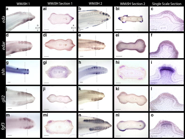
Fig. 4.

Gene expression analyses of early morphogenesis of caudal denticles. Expression of eda and its receptor edar are observed in the epithelium during early placode morphogenesis (a–f). eda can also be seen in tissue undergoing mineralisation later in morphogenesis (b–bi). shh is first observed in the epithelium during early morphogenesis, before becoming restricted to the basal epithelium later in morphogenesis (g–i). gli2 is also seen in the epithelium early during placode formation (j–l). Expression of fgf3 is first seen in the epithelium, before moving to both the epithelium and mesenchyme later in placode morphogenesis (m–o). The dashed lines show where in the WMISH the section was taken. WMISH Section 1 represents a younger stage specimen than WMISH Section 2. For the WMISH, D dorsal, V ventral, A anterior and P posterior. For WMISH sections, R right, L left, D dorsal and V ventral. For scale bars, a, b, d, e, g, h, j, k, m, n = 200 µm, ai, bi, di, ei, gi, hi, ji, ki, mi, ni = 100 µm, and c, f, i, l, o = 50 µm
