Fig. 1.

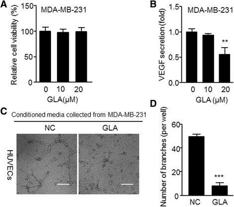
GLA reduces the angiogenic capacity in MDA-MB-231 cells. a MDA-MB-231 cells were exposed to 10 or 20 μM GLA for 48 h, using a Cell Counting Kit-8 assay of the cell vitalities, The percentage of cell viability was calculated via comparing with non-treated cells (mean ± SD, n = 3). Cells were then exposed to 10 or 20 μM GLA for 48 h, and conditioned media was collected. b The ELISA was used to detect the effects of GLA on VEGF secretion (mean ± SD, n = 3). MDA-MB-231 cells were pretreated with 0 or 20 μM GLA for 48 h, then the previous media was removed, and cells were washed with 1× PBS to replace fresh media with 1% serum for 24 h. The conditioned media was collected and incubated in (c) tube formation assays of the angiogenic capacity in MDA-MB-231 cells, HUVECs were exposed to the conditioned mediums collected as described in (b) for 6 h. d Quantitative analyses of the tube numbers, the total number of formed tube branches in each well was counted under the light microscope (mean ± SD, n = 5); ** P < 0.01 and *** P < 0.001 compared with cells treated without GLA
