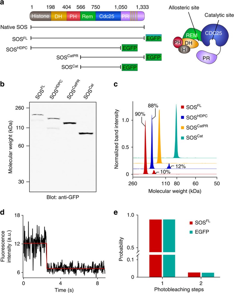
Figure 1. Characterization of various SOS constructs in HEK293T cell lysate.
(a) Domain organization of SOS. Native SOS and various EGFP-fused SOS constructs are shown. (b) Anti-GFP western blotting of SOS constructs from lysates. (c) Intensity profiles of western blot bands. Approximately 90% of SOSFL and SOSHDPC were expressed at the correct molecular weight. SOSCat and SOSCatPR were expressed as pure species. (d) Representative single-step photobleaching trace of SOSFL. (e) Histogram of photobleaching steps for SOSFL from lysate compared with purified, monomeric EGFP. SOSFL predominantly exists in a monomeric state. N is ∼200 for each protein.

