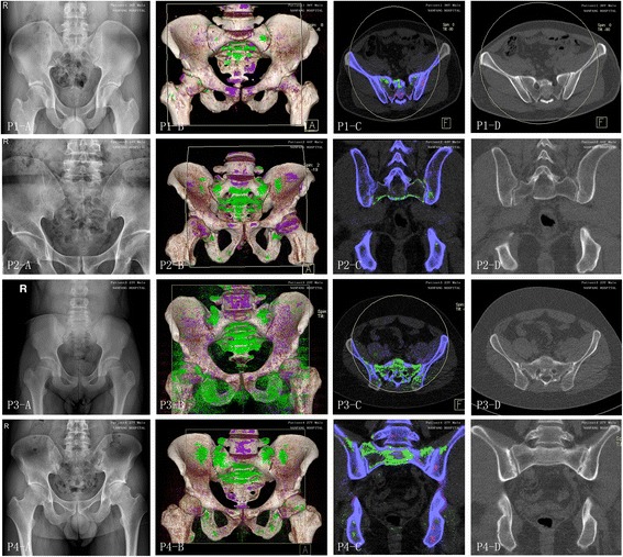
Fig. 2.

Four axial spondyloarthritis (AxSpA) patients with monosodium urate (MSU) crystal and radiographic structural damage at the sacroiliac joint. For each set of images, panel a shows the sacroiliac joint on plain radiographs, panel b shows the three-dimensional reconstruction dual-energy computed tomography (DECT) images, panel c shows the corresponding coronal (patient 2 and 4) or axial (patient 1 and 3) DECT images, and panel d shows the corresponding level of computed tomography (CT) images. A large quantity of MSU crystal deposition shown as green was found at the sacroiliac joint or the surrounding area in the DECT images. Four male patients (patient 1–4), aged 36, 44, 23, and 27 years old, had serum uric acid levels of 407 μmol/L, 370 μmol/L, 572 μmol/L, and 464 μmol/L, respectively. They were graded with a scale of 0, 0, II, III at the left sacroiliac joint and 0, I, II, III at the right sacroiliac joint, respectively, on plain radiographs
