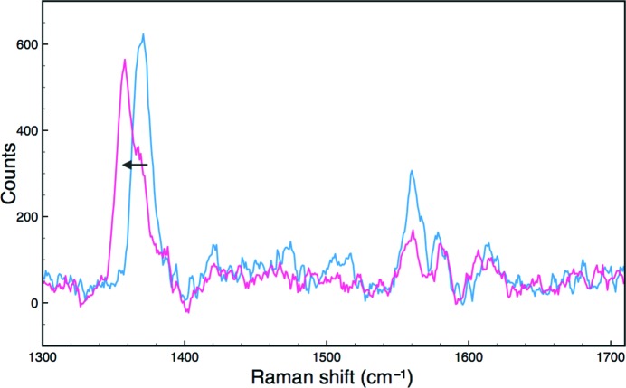
Figure 4.
High-frequency single-crystal resonance Raman spectra of DtpA at 100 K. Blue, ferric crystal spectrum before exposure to the X-ray beam; magenta, spectrum of the same crystal after collection of a diffraction data set. A shift in the ν4 redox-state marker band (arrow) is consistent with reduction to the ferrous form during data collection.

